Dr. Pradeep Kumar

Limb Preservation Surgery

Limb Preservation Surgery

1. Pre-operative Planning

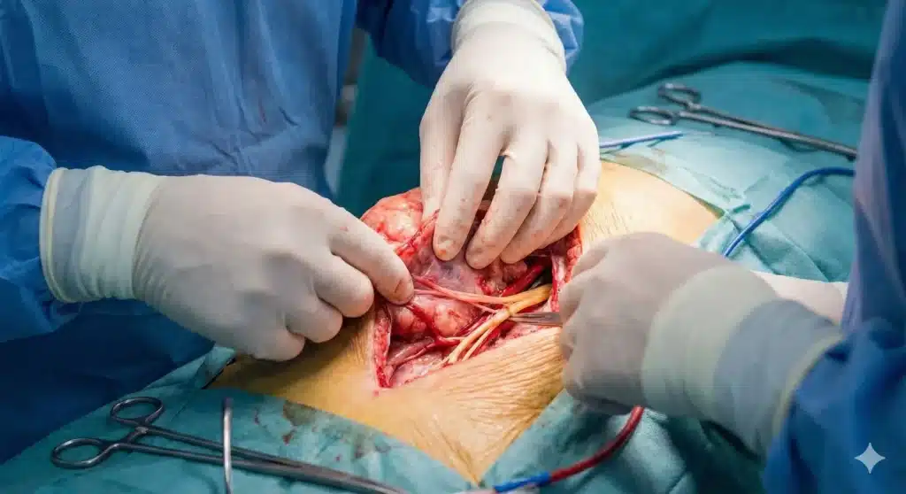
This image shows a surgeon meticulously planning the procedure using a high-resolution MRI scan. The scan highlights the location of the tumor and its proximity to vital structures, which is crucial for a successful limb-sparing operation.

2. Surgical Dissection and Nerve Preservation

This intra-operative photograph demonstrates the most critical part of the surgery. The surgeon is carefully dissecting the major neurovascular bundle (nerves and blood vessels) away from the tumor. This meticulous process ensures the limb’s function is preserved while removing the cancer with an adequate margin.

3. Post-operative Reconstruction

After the tumor has been successfully removed, the bone is reconstructed. This post-operative X-ray image shows the implantation of a modular prosthetic system. The implant replaces the removed distal femur and reconstructs the knee joint, restoring skeletal integrity.

4. Functional Recovery

The final image in the series depicts the ultimate goal of limb preservation: restoring function. It shows a patient in rehabilitation, walking with the help of a crutch and a physical therapist. The patient is wearing a knee brace and is actively working towards regaining mobility, demonstrating the successful outcome of the surgery.